Treatment of Macular Pucker in Baltimore
Also serving Pikesville, Owings Mills, Glen Burnie, & Rosedale
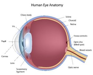
 The center of the eye is filled with a clear, jelly-like substance called vitreous. With age, the vitreous can shrink and pull away from the retina, or the layer of light-sensitive tissue lining the back wall of the eye. In some people, the vitreous tugs at the retina as it pulls away from it. As a result, scar tissue can develop and cause the retina to wrinkle or bulge. This is known as an epiretinal membrane. When an epiretinal membrane affects the macula, or the central portion of the retina responsible for clear central vision and fine detail, it is called a macular pucker.
Some macular puckers are asymptomatic. Others affect central vision, while peripheral (side) vision remains unaffected. Objects in the central field of vision can look wavy or small details may be difficult to make out. Gray or cloudy areas or blank spots can appear in the central visual field.
As renowned specialists in retinal diseases, Drs. Elman and Schechet are uniquely qualified to diagnose and treat macular puckers and restore clear central vision. The specific treatment for macular pucker depends on the symptoms and severity of the problem.
Vitrectomy
 Some macular puckers do not have a significant effect on vision and are monitored closely. If a macular pucker has a deleterious effect on vision, a surgical procedure called vitrectomy is the best option to correct the problem. The objective of vitrectomy surgery is to remove the eye’s natural vitreous and replace it with a saline solution. During the surgery, scar tissue is carefully removed to smooth out the pucker or wrinkle affecting the macula. This type of repair requires meticulous technique and the experience of qualified retinal surgeons like Dr. Elman and Dr. Schechet.
Surgical Details
Vitrectomy is normally performed on an outpatient basis. Patients are given an anesthetic around the eye and put into a “twilight sleep” state so they cannot feel any pain during the operation.
Our doctors begin the operation by creating three small, self-sealing incisions in the eye. They carefully remove the jelly-like vitreous, which allows them better access to the structures at the back of the eye. Our surgeons remove scar tissue and vitreous tugging on the macula to repair the wrinkling and help the macula return to its proper position.
During the surgery, Drs. Elman and Schechet replace the vitreous with balanced salt solution. Over time, the saline is replaced with the eye’s own fluid.
Recovering from Vitrectomy
After surgery, patients may experience temporarily reduced vision or the sensation that something is in the eye. Severe pain or other complications are very uncommon. With today’s advances in techniques and technology, vitrectomy normally yields very good outcomes.
Eyedrops and Ocular Medication
Eyedrops and other ocular medications cannot repair a macular pucker, but sometimes they are prescribed to help reduce associated swelling or leakage in the retina.
Learn More about Treatment for Macular Pucker
If you have been diagnosed with a macular pucker and are looking for qualified retinal specialists with whom to discuss treatment, Dr. Michael Elman and Dr. Sidney Schechet can help. Please schedule a consultation with them today to discuss your case and the available treatment solutions.
Macular Pucker FAQs
How is a Macular Pucker Diagnosed?
Our retinal specialists diagnose macular puckers during your comprehensive dilated eye exam, which includes imaging tests of the eye. After dilating your pupils with eye drops, Dr. Elman or Dr. Schechet will perform a retinal examination. If our doctors diagnose macular pucker, they will measure your visual acuity and check for blind spots or distortions in your vision using an Amsler grid. An Amsler grid is a block of intersecting vertical and horizontal lines that helps identify vision impairment.
What Are My Treatment Options for a Macular Pucker?
 A macular pucker often does not even require treatment because many macular puckers don’t cause any symptoms. Even with mild symptoms, many people adjust to everyday life with minor vision distortions caused by macular puckers. However, if wavy lines, blurry vision and other concerns impact your quality of life, eye surgery may be indicated to remove the macular pucker in order to improve your vision. The vitrectomy procedure reduces and prevents pulling on the retina by removing vitreous gel and replacing it with a sterile solution. The vitreous fluid in your eye is mostly water, so you won’t be able to tell the difference. The surgery addresses the pucker or wrinkle in the macula by removing the scar tissue.
Vitrectomy requires skilled hands and precision to improve your vision, but your entire field of vision may not return to normal. On average, about half of vision loss from macular pucker is restored and does so slowly over six to 24 months. Many of our patients see significant vision improvement after macular pucker treatment with vitrectomy, and this improvement continues month by month over one to two years.
How is Macular Pucker different from Macular Hole and Macular Degeneration?
The symptoms of these eye conditions are similar, but they are different disorders that require specific treatments to restore eyesight or prevent further vision loss.
Age-Related macular degeneration (AMD) occurs when the natural aging process damages the macula, causing vision loss in older adults. While it does not lead to complete blindness, the loss of central vision makes it challenging to see the faces of loved ones, read, drive and see fine details in general. AMD may develop slowly or quickly, depending on the form.
A macular hole may be caused by shrinking vitreous fluid when the vitreous fibers pull too hard on the retina and tear the central tissue (“fovea”) off of the macula, creating a macular hole. When this pulling causes damage to the retina’s surface and your body responds by healing and forming scar tissue, this is called macular pucker. In rare cases, a macular pucker may progress to a macular hole.
What Causes Macular Pucker?
The interior of your eye is mainly made up of vitreous fluid (about 80 percent), a gel-like substance that maintains the round structure of the eye. Millions of fine fibers in the vitreous attach to the retina. As you get older, the amount of vitreous decreases, and those fibers pull away from the retina, creating microscopic damage to its surface. Your retina heals, forming scar tissue, and when the scar tissue contracts, it creates a wrinkle or pucker. When this trauma and scar tissue occurs on the retina’s center (the macula), it affects your central vision. Previous eye surgery or injury may also cause macular pucker.
What are the Symptoms of Macular Pucker?
The severity of macular pucker symptoms varies from no vision disturbances to severe visual impairment. Typically, you will notice mild distortions such as straight lines appearing wavy or blurred vision. You may find it difficult to read fine print or small details and develop a blind spot or a gray area in your central field of vision.
Who is at Risk of Macular Pucker?
Most people who experience macular pucker are over 50 because it is an age-related concern. However, certain eye diseases may increase your risks, such as a detached retina and eye inflammation. People with diabetes are more at risk because diabetic retinopathy can lead to macular pucker. It may occur in one eye or affect the other eye later on as your vitreous fluid continues to shrink.