Retinal Vein Occlusion: When Blood Flow Affects Your Vision
Submitted by Michael J. Elman, M.D. on March 15, 2026
Retinal vein occlusion (RVO) is a serious vascular condition that can suddenly disrupt vision. It occurs when a vein responsible for draining blood from the retina becomes blocked, leading to swelling, bleeding, and reduced oxygen supply to retinal tissue. Because the retina plays a critical role in converting light into visual signals, any interruption in blood flow can significantly affect sight.
At Elman Retina Group, our board-certified retina specialists diagnose and manage retinal vein occlusion using advanced imaging technology and evidence-based treatment strategies. Early intervention is essential to preserving long-term vision.
What Is Retinal Vein Occlusion?
Retinal vein occlusion is often compared to a “stroke of the eye.” When a vein becomes blocked, blood and fluid back up into the retina, causing swelling. This is especially true in the macula, the area responsible for central vision. There are two primary types:
- Branch Retinal Vein Occlusion (BRVO): A blockage in one of the smaller retinal veins.
- Central Retinal Vein Occlusion (CRVO): A blockage in the main retinal vein.
The severity of vision changes depends on which vein is affected and the extent of the swelling or bleeding.
Symptoms to Watch For
Retinal vein occlusion may develop suddenly or gradually. Common symptoms include blurred or distorted vision, dark spots, or a noticeable decrease in central vision in one eye. Some patients experience mild vision changes at first, while others notice a significant loss of clarity. Any sudden change in vision warrants prompt evaluation by a retina specialist.
Risk Factors and Underlying Conditions
RVO is closely linked to systemic vascular conditions. Patients with high blood pressure, diabetes, high cholesterol, or glaucoma are at increased risk. Age is also a factor, as blood vessels naturally stiffen over time. Because retinal health reflects overall vascular health, managing these conditions is important for prevention and long-term care.
How Retinal Vein Occlusion Is Diagnosed
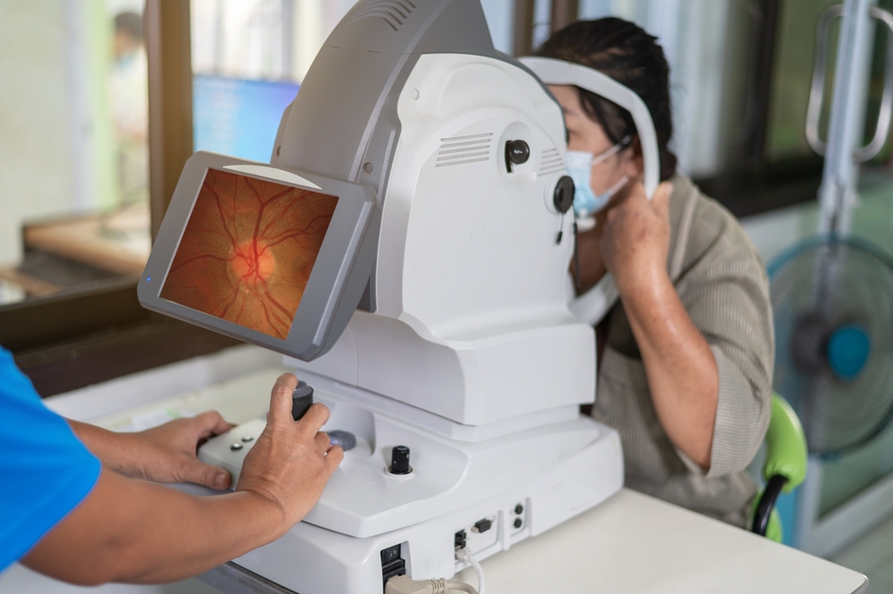
At Elman Retina Group, diagnosis involves a comprehensive dilated eye exam and advanced imaging. Optical coherence tomography (OCT) measures retinal swelling, while fluorescein angiography evaluates blood flow and identifies areas of leakage or blockage. These tools allow our specialists to determine the type and severity of the occlusion and guide treatment decisions.
Treatment Options and Prognosis
Treatment focuses on reducing macular swelling and preventing complications. Our doctors often recommend anti-VEGF injections to decrease fluid buildup and stabilize vision. In some cases, laser therapy may be appropriate to manage abnormal blood vessels. With early treatment and ongoing monitoring, many patients maintain functional vision despite the condition.
Expert Retinal Care in Baltimore, MD
Retinal vein occlusion requires specialized expertise and careful follow-up. At Elman Retina Group, our retina doctors provide comprehensive care in Baltimore, MD, with additional locations in Pikesville and Glen Burnie. If you notice sudden vision changes, contact our team at (410) 686-3000 to schedule an evaluation and protect your sight.
We’re happy to hear from you! Please be aware that this form is not monitored closely and isn’t the best way to reach us with medical questions. For timely assistance, please call the office directly, and avoid sharing medical or sensitive information here.