Human Eye Anatomy
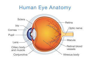 The experienced ophthalmologists at Elman Retina Group in Rosedale believe in patient education. Understanding your condition begins with knowing the various parts that comprise the human eye, and how an issue with any one of those parts can affect the other structures.
If you’re experiencing vision changes, eye pain or other eye-related concerns, schedule an eye exam with our board-certified ophthalmologists, Dr. Michael Elman and Dr. Sidney “Sid” Schechet.
Structures of the Human Eye
Our adventure into the eye’s anatomy starts in the extraocular muscles and journeys inward. Learn about the function of these critical components of your eye.
Extraocular Muscles
Six extraocular muscles attach to the eye’s sclera (white portion). These muscles allow your eye to move upward, downward and side to side, and to rotate. The levator palpebrae superioris is the triangular muscle inside the upper eyelid that allows for blinking. Some issues that may occur with these muscles include blepharospasm (uncontrollable blinking), strabismus (crossed eyes) and ptosis (drooping eyelid).
Tear Film
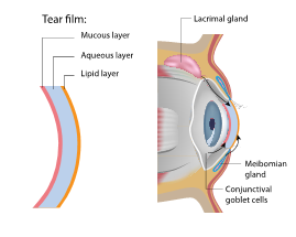 The tear film covering your eye’s surface is made of a delicate balance of mucous, water and oil. The meibomian glands on the inside of the upper and lower eyelids secrete the lipid (oil) layer of your tears. The lacrimal glands above each eyeball under the outer edge of the eyebrow produce the aqueous (water) layer, while conjunctival goblet cells create and spread the mucin (mucous) tear layer. These layers work together to protect your eyes and drain through the tear duct. Any disruption in tear creation, ratio or drainage can cause problems such as dry eye disease.
Sclera
The white section of the eye supports the wall of the eyeball, helping it keep its round shape and preventing injury. The sclera is a tough structure that stops debris from penetrating or rupturing the eye. Scleral problems are rare but may include scleritis (inflammation of the sclera).
Conjunctiva
The conjunctiva is the clear tissue that coats the sclera and the inside of the eyelids. It provides lubrication and protection by creating the mucus layer of the tear film and stops microbes from entering the eye. Eye problems with the conjunctiva include conjunctivitis (commonly known as pink eye). Conjunctival injury, such as overexposure to the sun, a foreign body in the eye, infection or allergic reaction can lead to a gritty or burning feeling, blurry vision and dry eye symptoms.
Cornea
The cornea is a clear, dome-shaped structure in the front of your eye that helps focus incoming light onto the retina. A mishappen cornea is the reason people have trouble seeing objects clearly and is the cause of nearsightedness, farsightedness and astigmatism. Vision correction procedures, such as LASIK, reshape the cornea to improve vision. Other common issues with the cornea include corneal ulcers, abrasions and keratitis.
Iris
The iris is the pigmented section of your eye that controls how dilated your pupil is at any given moment due to the pupillary light reflex. The muscle of the iris adjusts the circular opening (pupil) to regulate how much light is directed onto the retina. That muscle reflex is why you notice your pupils are small in bright sunlight and large in low-lit environments. Problems with the iris include iritis, or inflammation of the iris that causes light sensitivity, headache, eye pain and decreased vision.
Drainage Angle
The eye’s drainage angle is where the aqueous humor (clear liquid inside the front portion of the eye) is funneled out from the eye to maintain proper intraocular pressure. It’s found between the cornea and iris, where the iris and sclera meet. Issues with the drainage angle cause a group of eye diseases called glaucoma. Glaucomas may be open-angle or closed-angle and are one of the most severe and common types of eye diseases.
Lens
The lens of your eye is located behind the iris and works with the cornea to refract (bend) light onto the retina for vision. Just like the lens of a camera, the eye’s natural lens changes shape to focus on objects at various focal distances. Eye problems with the lens include cataracts, a leading cause of blindness but also an eye disease that is curable through cataract surgery. Proteins in the eye clump together and cloud the eye lens, causing muted and blurry eyesight. The lens is removed and replaced with an artificial lens implant.
Another common concern with the lens is presbyopia, which occurs as you age and the lens loses its flexibility, causing blurry near vision. Presbyopia is why adults over 40 require reading glasses.
Retina
The light-sensitive nerve tissue lining the back wall inside your eye is called the retina, and it is a critical factor in your ability to see. The cornea and lens focus light onto the retina, which sends light messages to your brain through the optic nerve. There are many potential concerns with the retina, including retinal detachment, diabetic retinopathy and proliferative vitreoretinopathy. Retinal problems often require timely treatment with an ophthalmologist to preserve your vision.
Macula
 Located at the center of the retina, the macula is a critical anatomical feature of the eye, providing central vision so you can view people and objects directly in front of you. Age-related macular degeneration is one of the biggest concerns relating to the macula because this eye disease thins the macular tissue, creating blurry or splotchy central vision that can’t be restored, only stopped. Other issues with the macula include macular edema, macular holes and macular puckers.
Aqueous Humor
The aqueous humor is a clear liquid in the front of the eye between the lens and cornea that nourishes and cleans the lens and balances the pressure inside the eye. Aqueous humor drains waste from the eye via the drainage angle. A build-up of aqueous humor that does not drain correctly can cause damage to the optic nerve as part of glaucoma.
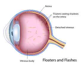
Vitreous Humor
Vitreous humor is a transparent, gel-like substance in the rear portion of the eye between the lens and retina that supports the retina and provides nutrients to the lens. The gelatinous vitreous can detach, causing floaters and flashes of light in your side vision.
Schedule Your Eye Exam at Elman Retina Group
Our ophthalmologists are board-certified by the American Board of Ophthalmology and are leading experts in various eye conditions, diseases and traumas. If you’re experiencing eye-related concerns, schedule an eye exam at our Rosedale office.
Contact Elman Retina Group by calling (410) 686-3000 or filling out our online contact form.
We’re happy to hear from you! Please be aware that this form is not monitored closely and isn’t the best way to reach us with medical questions. For timely assistance, please call the office directly, and avoid sharing medical or sensitive information here.