Central and Branch Retinal Vein Occlusion
Also serving Pikesville, Catonsville, Glen Burnie, & Rosedale
Central and Branch Retinal Vein Occlusion
 Your eye’s retina relies on good blood supply to receive the oxygen and nutrients it needs to function properly. Retinal arteries carry blood from your heart to your retina, and retinal veins carry the blood from your retina back to your heart.
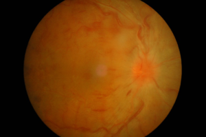
Retinal vein occlusion (RVO) is a condition in which the tiny veins of the retina become obstructed. Central retinal vein occlusion is a blockage of the main retinal vein, and branch retinal vein occlusion is a blockage of one of the smaller branches of this main vein. Both types of RVO affect your ability to see clearly.
When a blockage occurs in the central retinal vein, its effects are felt immediately. As circulation slows, the center of the retina experiences an accumulation of blood and fluid. This backup interferes with the retina’s function of receiving images. Blurry vision and other impairments may result.
With branch retinal vein occlusion, the blockage of a branch vein also creates macular edema, but the reduction in blood flow (ischemia) may be less, as other veins and capillaries continue to carry blood through the retina. This reduces visual acuity, but the effects may be less severe or sudden than a blockage of the central retinal vein.
If you develop a complication like retinal vein occlusion, prompt attention is needed. The trusted team of doctors at Baltimore-area Elman Retina Group have the knowledge and skills to determine the most suitable treatment and restore your retina to good health.
Retinal Vein Occlusion FAQs
What role does high blood pressure play in the development of RVO?
Just as high blood pressure is harmful to other areas of your body, it is also damaging to the veins and capillaries that keep your retina supplied with blood. High blood pressure is also associated with the development of atherosclerosis. Because the “hardened” arteries of atherosclerosis are heavier and less flexible than normal, the main retinal artery can begin to put pressure on the surrounding veins and capillaries, reducing overall flow.
Can RVO be treated in one office visit?
Once you are diagnosed with RVO, you will need to begin ongoing treatment that will allow your Elman Retina Group ophthalmologist to monitor the progression of the disease and administer regular injections to relieve pressure. Neovascular glaucoma develops in about a third of RVO cases, resulting in an unsafe rise in eye pressure that puts your vision at risk.
Are there other possible complications?
Your ophthalmologist also needs to check regularly for any signs of hemorrhaging. One of the consequences of high eye pressure levels is pressure on the small capillaries within the eye. They may begin to leak, and the resulting diffusion of blood within the eye can partially cover photoreceptors. When this happens, light and images cannot reach the photoreceptors, and your vision is impaired. This can be temporary, or in some cases a chronic condition that can persist for years.
In some cases of branch RVO, retinal neovascularization appears on the surface of the retina. This is an abnormal growth of new capillaries which may be prone to rupture and hemorrhaging.
Will RVO change the way I live?
One important change you will need to make in your lifestyle is to be very careful when performing activities that require accurate depth perception. Blurred vision in the affected eye will reduce the brain’s ability to process and judge distances accurately. This can affect your dexterity in tasks small and large — everything from pouring hot coffee into a cup without a spill to driving a car without an incident. If your work involves working with heavy machinery or climbing ladders, this may no longer be practical or safe.
Schedule a Consultation with the Team at Elman Retina Group
To discuss any changes to your vision with the doctors at Elman Retina Group, please request an appointment with us today.