3 Parts of Your Eye Anatomy That Make Vision Possible
Submitted by Elman Retina Group on December 6, 2023
Intricate eye structures work together to make your vision possible, transforming incoming light into images. How clearly you see those images depends on several parts of your eye, and it all comes down to how your eye bends or refracts light. If the light is not focused properly onto the back of your eye, you will have blurry vision. Eye diseases and natural aging also affect these structures and how well you see. Below, our eye doctors at Elman Retina Group explain how your eyesight works and how to improve your vision.
1. Cornea
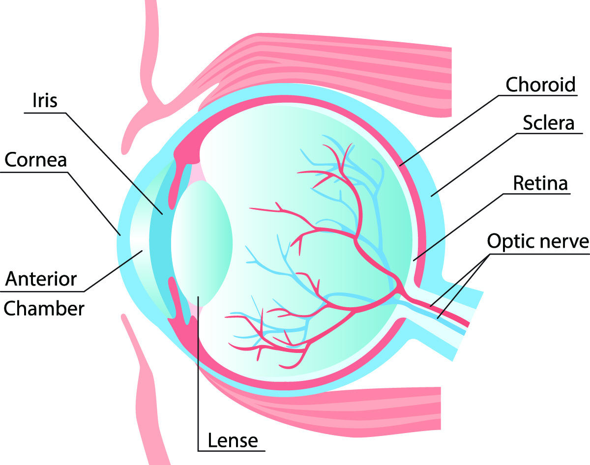
The cornea is the clear dome located on the front of your eye. Incoming light goes through the cornea and is directed through and onto other structures to create vision. If the cornea is misshapen, you will see blurry images without corrective eyewear, such as contact lenses and prescription eyeglasses. Astigmatism, nearsightedness, and farsightedness are refractive errors caused by an abnormally shaped cornea, but laser vision correction can correct these vision problems. Some eye conditions affect the corneal shape, such as keratoconus, which causes the cornea to bulge outward like a cone.
The light that enters the cornea goes through the iris and pupil. The iris is the colorful or pigmented part of your eye that controls pupil dilation with the pupillary light reflex. The iris contains muscles that adjust the circular pupil, allowing more or less light into the eye. You have likely noticed your pupil shrinking in bright sunlight and growing larger in low-lit places.
2. Lens
The cornea works with the lens to focus light rays onto the back of the eye where the retina lives. The lens sits behind the iris and adjusts focus for near, intermediate, and distant focal points. Presbyopia is a type of refractive error that happens with age when the lens becomes more rigid and has difficulty focusing on nearby objects. Cataracts develop in the lens as proteins break apart and clump together, creating a cloudy or opaque appearance. Cataract surgery and refractive lens exchange can restore clear vision from cataracts and presbyopia.
3. Retina
A light-sensitive tissue layer lines the back of the eye, called the retina. The retina receives light from the cornea focused by the lens, and photoreceptors in the retina turn the light messages into electrical signals that travel through the optic nerve to the brain. The brain turns the electrical signals into images. Where the light comes to a point affects how well you see. When the light meets an endpoint before the retina, you will have nearsightedness (myopia), and when it meets after the retina, you will have farsightedness (hyperopia). Some eye diseases or injuries affect the retina, such as retinal detachment, diabetic retinopathy, and proliferative vitreoretinopathy.
The middle of the retina is called the macula, and this area is responsible for your central vision. The macular may develop holes, puckers, or edema (swelling), causing vision loss or blindness. Age-related macular degeneration is a serious eye disease that causes gradual or progressive vision loss that can’t be restored, only stalled.
This pathway to vision can also be damaged by glaucoma. Glaucoma causes an increase in the internal eye pressure that damages the optic nerve, affecting the messages sent to the brain and causing loss of peripheral (side) vision first.
If you have blurry vision or are experiencing signs of eye disease, contact our eye doctors at Elman Retina Group at (410) 686-3000. Our board-certified ophthalmologists serve patients in Glen Burnie and Pikesville, Maryland.
We’re happy to hear from you! Please be aware that this form is not monitored closely and isn’t the best way to reach us with medical questions. For timely assistance, please call the office directly, and avoid sharing medical or sensitive information here.