How Much Do You Know About the Anatomy of Your Eye?
Submitted by Elman Retina Group on March 12, 2022
Despite relying on our eyes almost constantly throughout the day, most people are not that familiar with this complex part of the body. In fact, take a moment and ask yourself: How much do you really know about the anatomy of your eye?
Read on as Elman Retina Group’s doctors take a closer look at the many parts of the eye that work together to provide the extraordinary gift of sight.
Basic Anatomy of the Eye
The eye is surrounded by the orbit (a bony socket). Six extraocular muscles within the orbit control eye movement and alignment. The fastest-moving muscle in the body is the orbicularis oculi, which can close the eyelid in less than 100 milliseconds. This is why we use the phrase “in the blink of an eye” to describe an event that happens very quickly.
Tissue called the conjunctiva covers the white part of the eye and lines the inner eyelids. Most of us have experienced inflammation or irritation of the conjunctiva at one point or another (we call it by a casual term: “pink eye”).
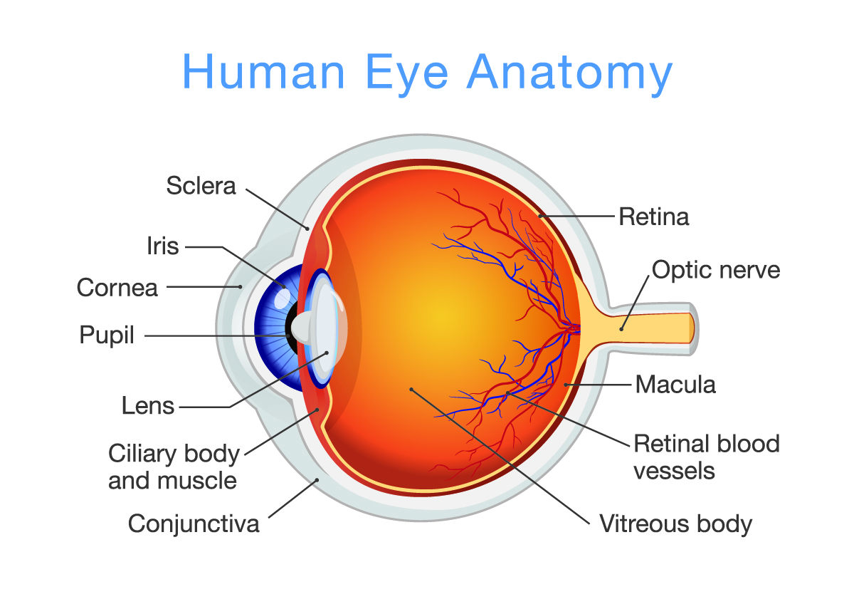
Anterior of the Eye
At the front of the eye is a dome-shaped covering called the cornea. The cornea is responsible for 70 percent of the eye’s focusing power. Immediately behind the cornea sits the anterior chamber, which is filled with a clear liquid called aqueous humor that nourishes the eye.
Behind the anterior chamber is the colored portion of the eye known as the iris. Incoming light passes through a small opening in the iris, called the pupil, on the way to the back of the eye. The pupil can widen (“dilate”) to allow more light into the eye or constrict to limit incoming light as needed.
The eye’s lens sits behind the iris. Responsible for the remaining 30 percent of the eye’s focusing power, the lens changes shape to focus on objects at different distances.
Posterior of the Eye
In between the lens and the back of the eye is a cavity containing vitreous humor, a gelatinous substance that helps maintain the eye’s spherical shape.
At the back of the eye sits the retina, which sends images to the brain by way of the optic nerve. You probably didn’t know that images are focused on the retina upside down and backward; the brain must turn the images right side up.
The retina has millions of special cells called rods and cones. Rods allow us to detect light and dark, and cones detect color. Colorblindness which disproportionately affects more men than women, occurs when one of the three kinds of cones responsible for bringing world into color are missing. The macula is a small part of the retina that enables clear central vision.
You cannot feel anything in your retina. Unlike problems affecting the skin and bones, retinal problems cause no physical sensations — so unless you stay carefully attuned to changes in your vision and see an eye doctor regularly, retinal problems are likely to go undetected.
Contact Our Retinal Specialists
If you have noticed your vision changing, book an eye exam with a qualified eye doctor to determine the underlying cause. Should your doctor suspect a disorder affecting your retina, our specialists at Elman Retina Group are ready and willing to help. Call us at 410-686-3000 or visit our Pikesville or Glen Burnie offices today!
We’re happy to hear from you! Please be aware that this form is not monitored closely and isn’t the best way to reach us with medical questions. For timely assistance, please call the office directly, and avoid sharing medical or sensitive information here.